
Microscopy and Biophysical Methods Laboratory
Methods at a Glance
- Micropipette video microscopy
Inverted digital video microscopy with micropipette manipulation to visualize and control single microdroplets and model membranes in real time.
- Confocal Raman microscopy
Confocal Raman spectroscopy/microscopy to obtain vibrational spectra from defined regions of lipid membranes and droplet interface bilayers, revealing molecular structure, composition, and membrane perturbations by bioactive molecules.
- Interfacial tensiometry
Rame‑Hart tensiometer with DROPimage analysis to measure liquid–gas and liquid–liquid interfacial tensions and quantify monolayer formation and surfactant/lipid adsorption.
- Electrophysiology
Custom setups to probe electrical properties of droplet interface bilayers and related membrane systems, including ion transport, membrane integrity, and the effects of membrane‑active compounds.
- ATR‑IR spectroscopy
Attenuated Total Reflectance IR for probing molecular structure and organization in lipid membranes and interfacial assemblies at soft interfaces.
- Differential scanning calorimetry (DSC)
TA Instruments DSC to characterize lipid phase transitions and quantify how interactions with drugs, cholesterol, and other bioactive molecules alter membrane thermodynamic properties.
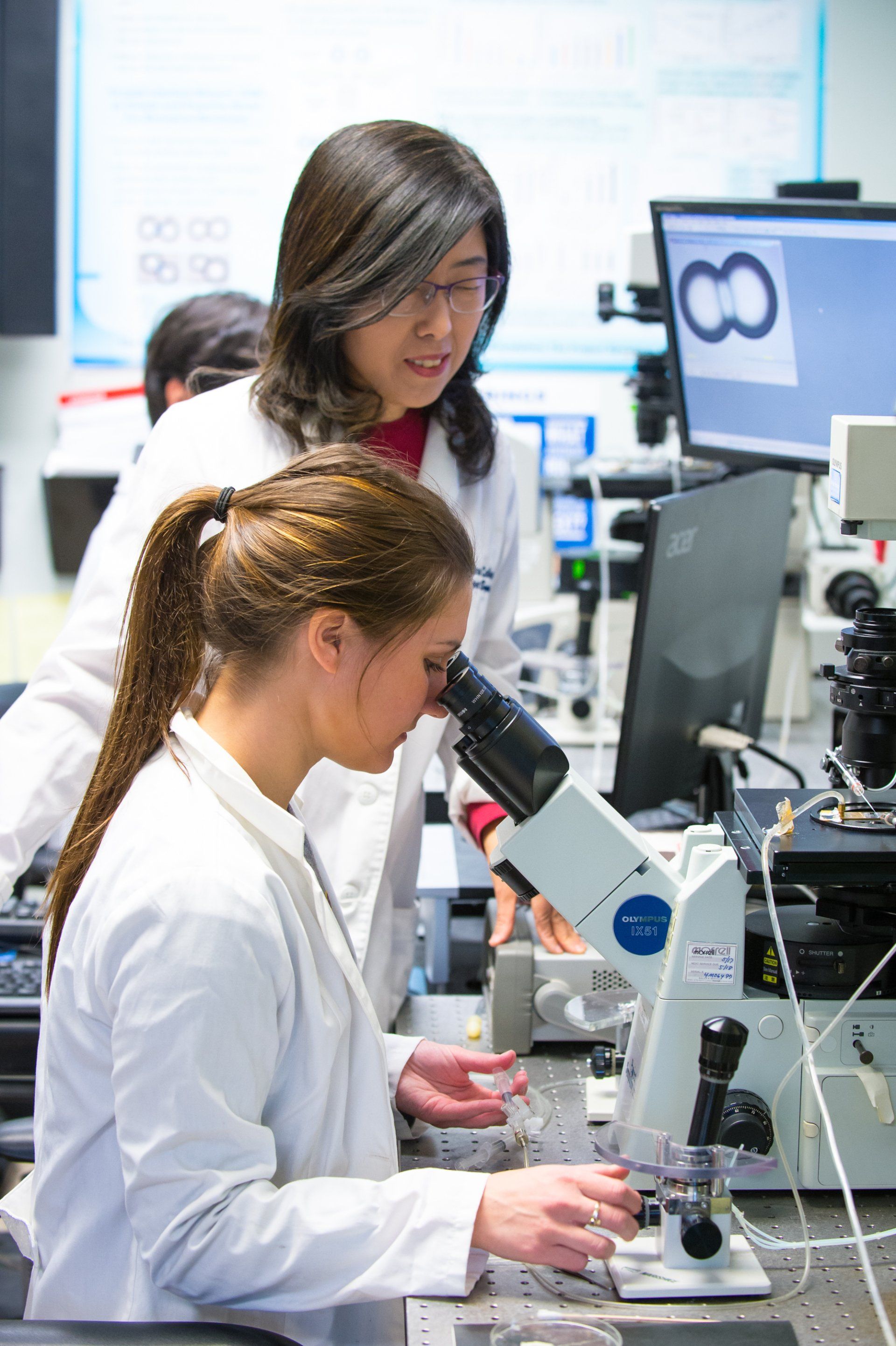
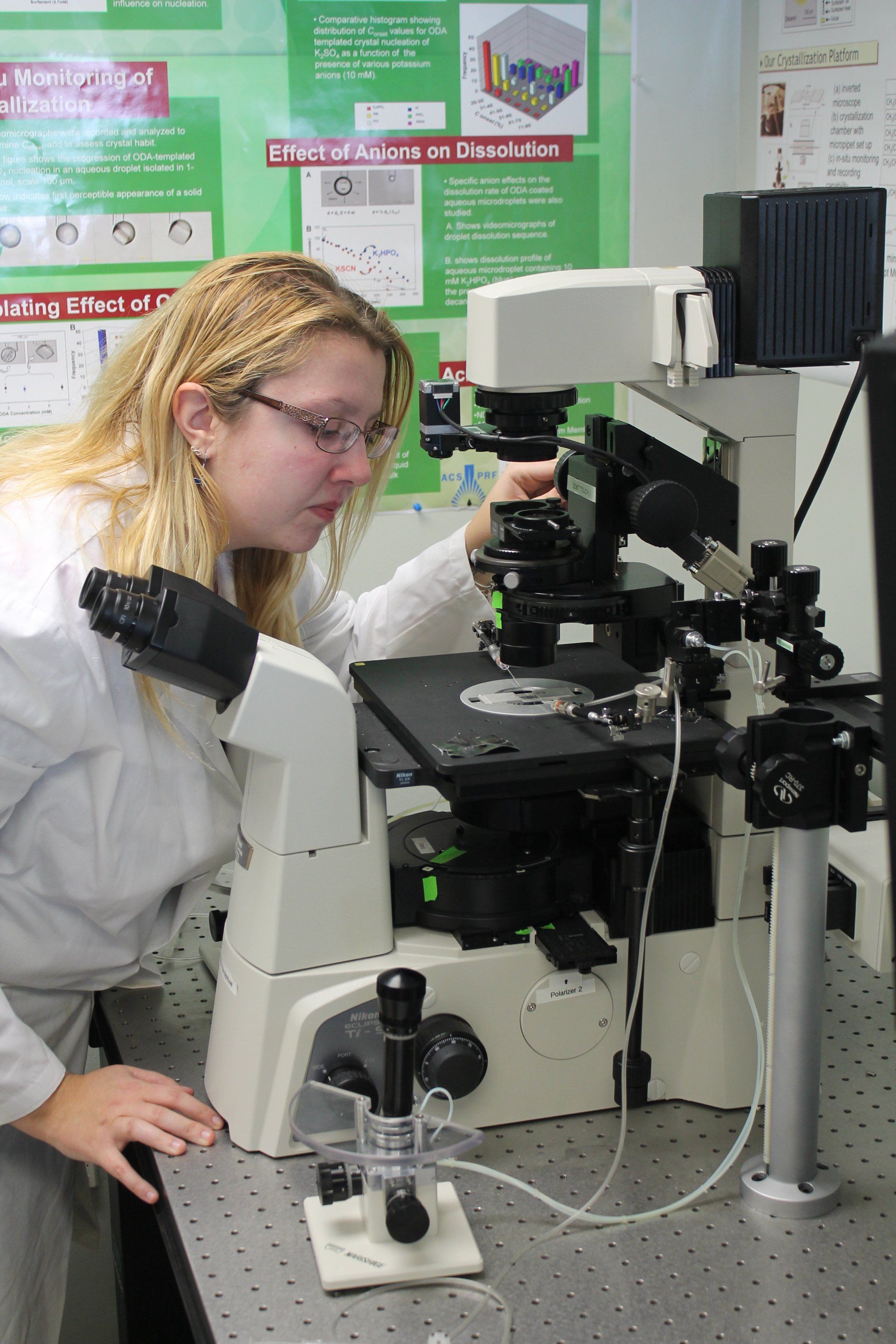
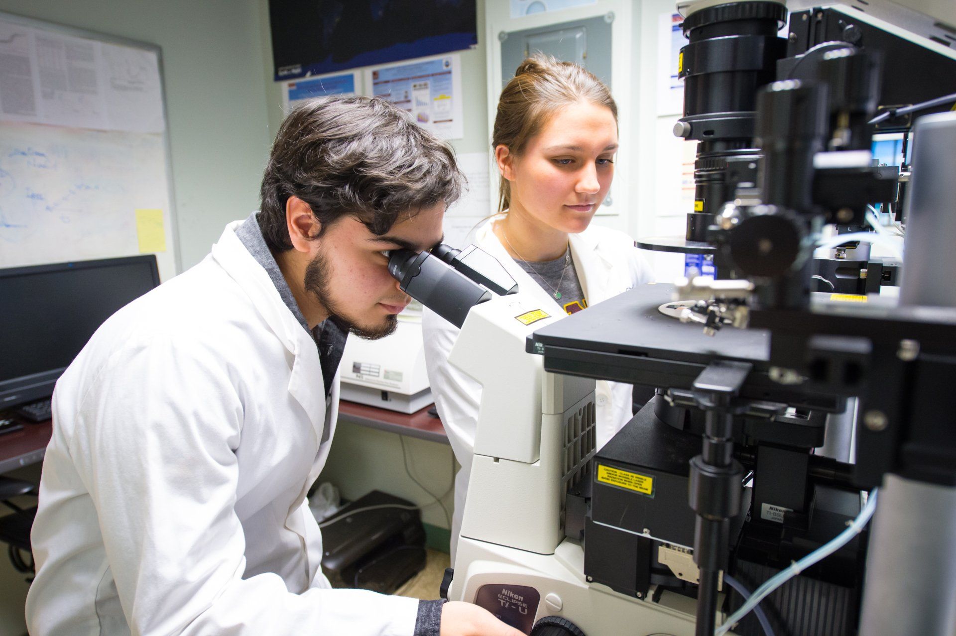
Micropipette Video Microscopy
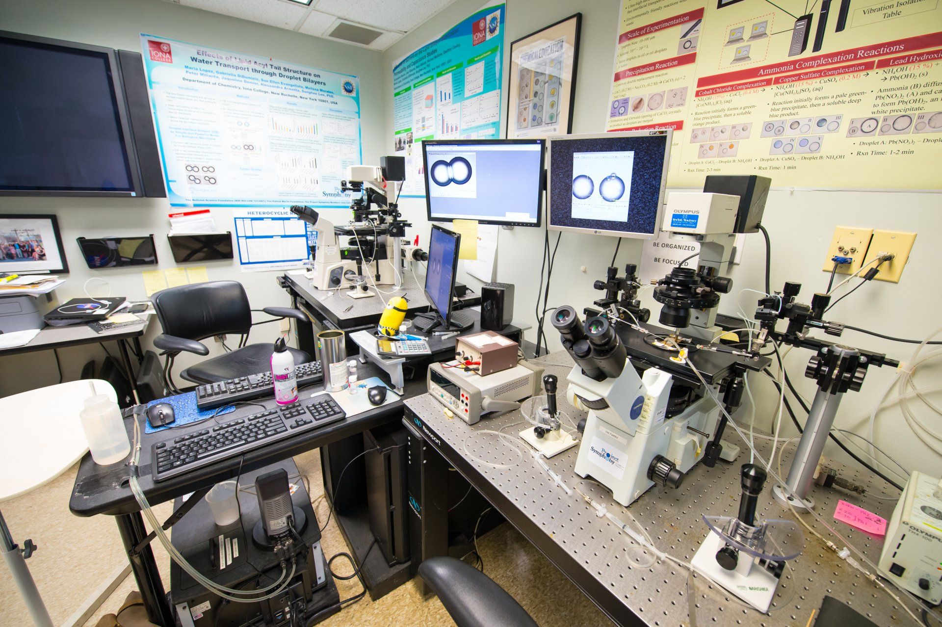
A core technique in our laboratory is inverted digital video microscopy combined with micropipette manipulation (micromanipulation). Our setup consists of an experimental station (inverted microscope with micromanipulators), a data acquisition station (digital camera and computer interface), a distribution system (digital library of images and videos), and a projection/display system. This platform allows us to visualize and record reactions between and within aqueous microdroplets in real time.
Micropipette Manipulators
Micropipette manipulators allow us to create, position, and manipulate single, cell‑sized droplets in a well‑defined and controlled liquid environment. By translating, merging, or separating droplets, we can change the surrounding environment, introduce reagents, and modulate interfacial monolayers and bilayers. These capabilities are central to our studies of membrane mimics, interfacial assembly, and microscale chemical phenomena.
Inverted Confocal Raman Microscope System (XploRA INV from Horiba)
Confocal Raman spectroscopy/microscopy to obtain vibrational spectra from defined regions of lipid membranes and droplet interface bilayers, revealing molecular structure, composition, and membrane perturbations by bioactive molecules. The Inverted Confocal Raman Microscope with micromanipulation system has two lasers consisting of 532 nm 25 mW solid state laser with spectral resolution, 0.7 cm-1/pixel and 785 nm 100 mW laser diode, with spectral resolution, 0.4 cm-1/pixel.
Attenuated Total Reflectance (ATR)‑FTIR spectroscopy
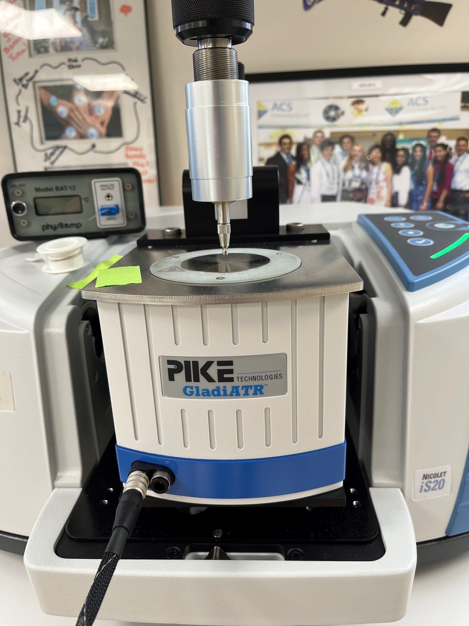
Attenuated Total Reflectance Fourier-Transform Infrared (ATR‑FTIR) spectroscopy is used to study the structure and organization of lipid membranes and interfacial assemblies. By monitoring vibrational bands associated with lipid headgroups, hydrocarbon chains, and interacting solutes, we gain molecular‑level insight into how bioactive molecules associate with membranes and alter their structure. These measurements are carried out using a Thermo Scientific Nicolet iS20 spectrometer equipped with a deuterated triglycine sulfate (DTGS) detector and a GladiATR single‑reflection ATR accessory with a diamond crystal (Pike Technologies, USA).
Differential Scanning Calorimeter (DSC, TA 2000)
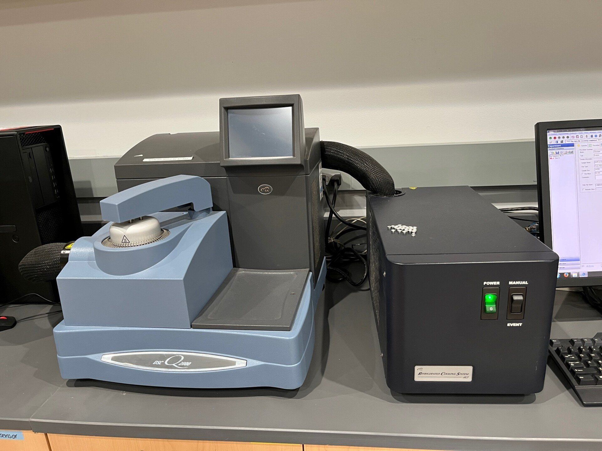
Differential Scanning Calorimetry (DSC; TA Instruments 2000) is a thermodynamic technique that measures heat flow associated with transitions in materials as a function of temperature and time. In our work, DSC is used to characterize lipid phase transitions and to quantify how interactions with exogenous molecules—such as drugs, cholesterol, or other bioactive compounds—shift membrane transition temperatures and enthalpies. These measurements help link molecular interactions to changes in membrane physical properties.
Interfacial tension measurements (Rame-Hart Goniometer/Tensiometer)
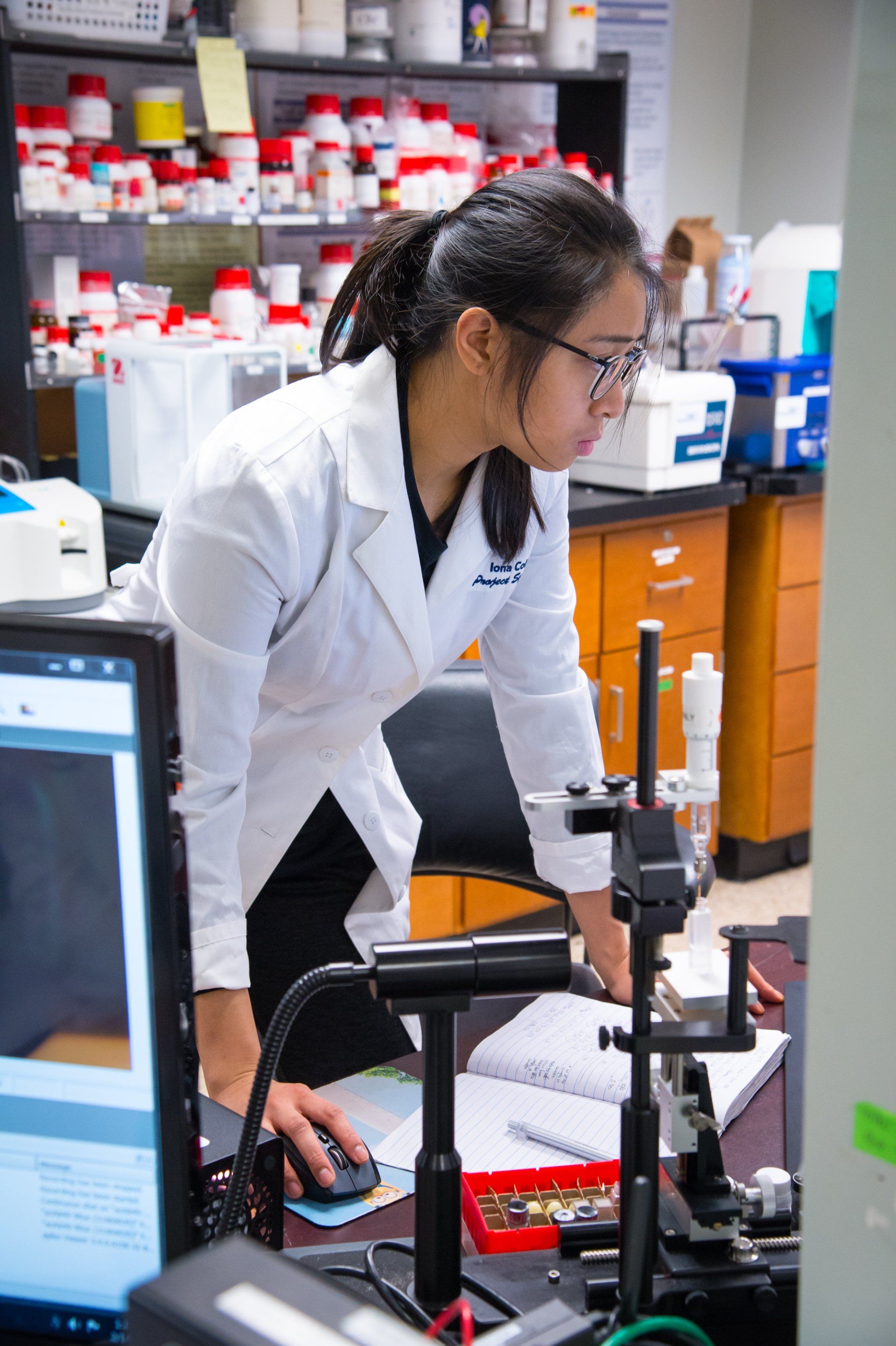
The study of how surfactants spread at liquid-liquid interfaces can lead to a better understanding of monolayer formation in colloidal systems. One direct measure of this transfer is the change in the interfacial (liquid-gas and liquid-liquid) tension. The interfacial tension of water against a solution of lipid in oil will allow us to determine area per molecule of the lipid at the interface, using the Gibbs adsorption equation, therefore, provide an evidence of interfacial densities. A Rame-Hart Tensiometer is used in conjunction with image analysis software DROPImage for measurement of interfacial tension.
Electrophysiology (Custom-made)
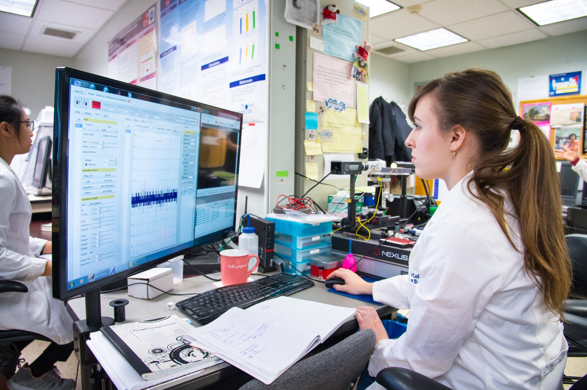
We also employ custom‑built electrophysiology setups to probe electrical properties of droplet interface bilayers and related membrane systems. These measurements provide complementary information about ion transport, membrane integrity, and the effects of bioactive molecules on membrane conductance and barrier function.